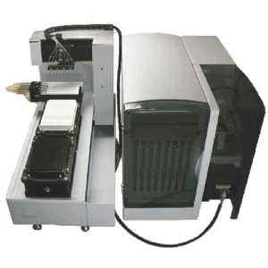


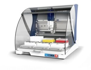


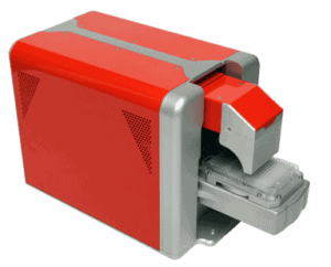

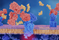


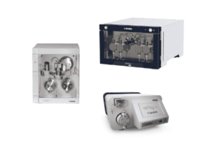


Next-generation label-free analysis
BLI is a powerful technology that is flexible, highly-sensitive and accessible. From experiment to analysis, characterise the widest range of molecules on a single easy-to-use platform ― in a single run.
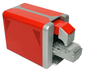
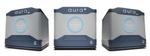
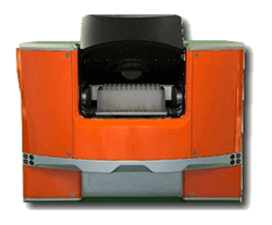
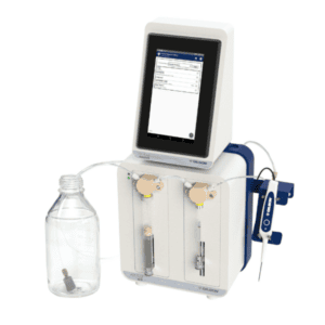
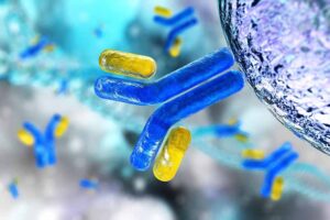
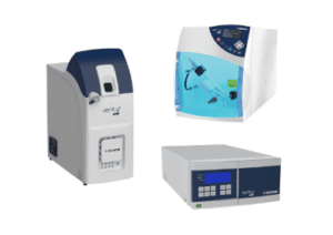
True low volume, high throughput subvisible particle analysis
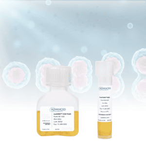
Aura systems help you develop stable, pure, and efficacious protein, cell, and gene therapies faster. By combining the sensitivity of BMI with fluorescent detection, Aura systems enable high-throughput particle count, size and identification.
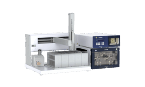
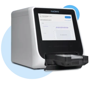
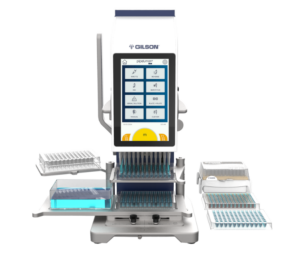
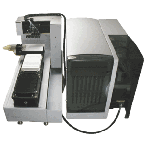
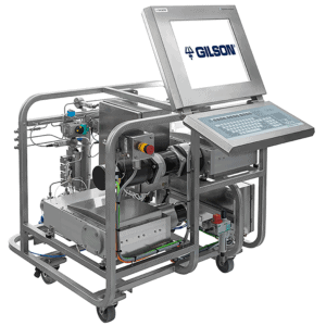
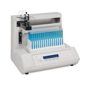
Rapid protein prototyping – Express protein on your own terms
Nuclera’s eProtein Discovery is a benchtop system combining digital microfluidics alongside in situ protein detection assays and cell-free protein synthesis. The eProtein Discovery™ solution, including integrated eProtein Discovery Software, gets protein in your hand quicker than ever before. With our cell-free protein synthesis reagents and microfluidics-based eProtein Discovery™ cartridges, it all happens on your own benchtop, in your way.
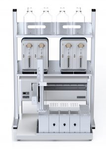
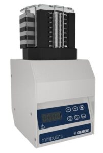
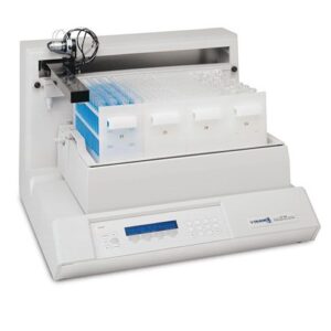
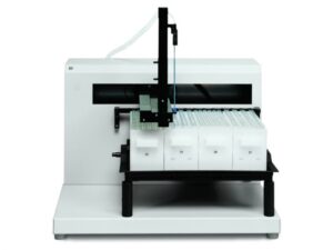
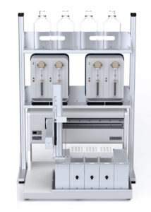
Advancing bioprocessing with precision & performance
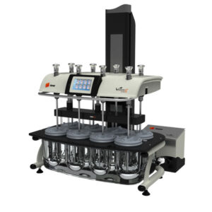
Explore Sepax’s portfolio where innovation meets reliability. Empower your processes with cutting-edge solutions designed for excellence in biomolecule separation and purification.
Next-generation label-free analysis
BLI is a powerful technology that is flexible, highly-sensitive and accessible. From experiment to analysis, characterise the widest range of molecules on a single easy-to-use platform ― in a single run.
True low volume, high throughput subvisible particle analysis
Aura systems help you develop stable, pure, and efficacious protein, cell, and gene therapies faster. By combining the sensitivity of BMI with fluorescent detection, Aura systems enable high-throughput particle count, size and identification.
Rapid protein prototyping – Express protein on your own terms
Nuclera’s eProtein Discovery is a benchtop system combining digital microfluidics alongside in situ protein detection assays and cell-free protein synthesis. The eProtein Discovery™ solution, including integrated eProtein Discovery Software, gets protein in your hand quicker than ever before. With our cell-free protein synthesis reagents and microfluidics-based eProtein Discovery™ cartridges, it all happens on your own benchtop, in your way.
Advancing bioprocessing with precision & performance
Explore Sepax’s portfolio where innovation meets reliability. Empower your processes with cutting-edge solutions designed for excellence in biomolecule separation and purification.
Countstar’s Castor X1 and Castor S1 systems are high-throughput image analysers that enable high-content analysis of thousands of cells simultaneously. Castor analysers are equipped with advanced optical technologies and Artificial Intelligence (AI) based image recognition algorithms, ensuring high quality images and accurate image processing for confident results.
Castor analysers are equipped with advanced optical technology, allowing the capture of clear and sharp images in the entire well area, ensuring accuracy. Castor analysers run on Peltier-cooled 10MP CMOS high resolution camera, 4x and 10x high NA objectives and a laser-based auto-focus system, ensuring sharp image acquisition in any situation.
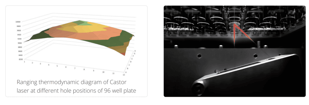
Differences in focal planes of single wells on a microtitre plate, either due to the specific design or due to variations in production batches, lead to inconsistent image quality. Castor analysers scan each well bottom with a laser to determine the optimum focus in milli-seconds, thus ensuring clear images of each well. The laser ranging auto-focusing also allows the usage of multi-well plates from a wide range of suppliers.
The concave meniscus formed in the wells of a microtitre plate causes significant light refraction near the edge of a well, resulting in an image with dark ring section when using common imaging technology. This makes it challenging to identify and quantify cells seeded near the edges. Castor's innovative imaging technology solves this issue, captures bright and clear images of the entire well area, enabling confident identification of single cells near the well’s edge.
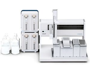
Castor analysers contain all the necessary components, interfaces, and software modules to be integrated into various automated platforms, from simple multi-axis robotic arm with plate stackers to fully automated station with sample preparation and liquid handing stations. Built-in barcode readers guarantee secure and traceable identification of each sample carrier.
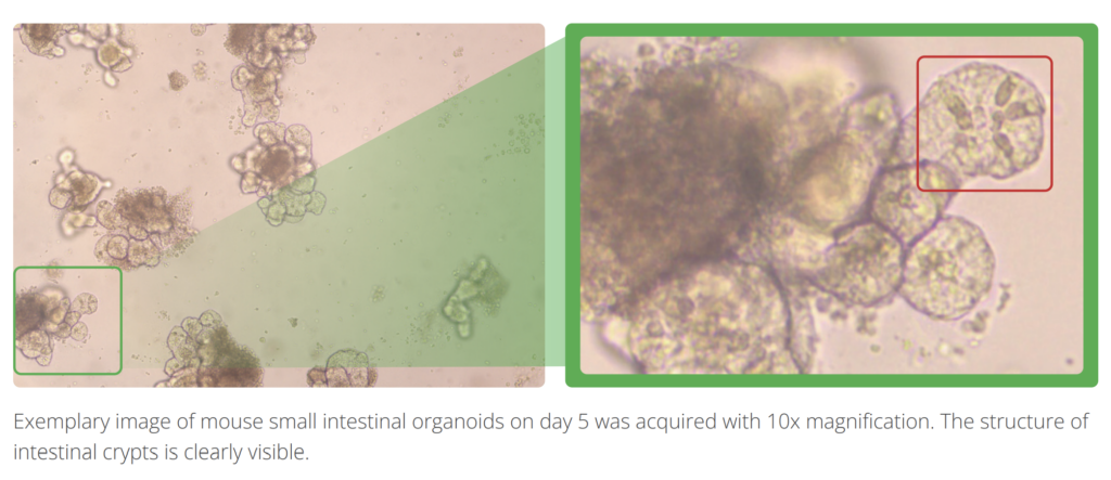
Castor analysers are equipped with a high-sensitivity, Peltier-cooled CMOS camera with a resolution of 10MP. The 4x and 10x objectives of high numerical aperture (NA) captures high-resolution images which enables precise phenotyping of cells and organoids.
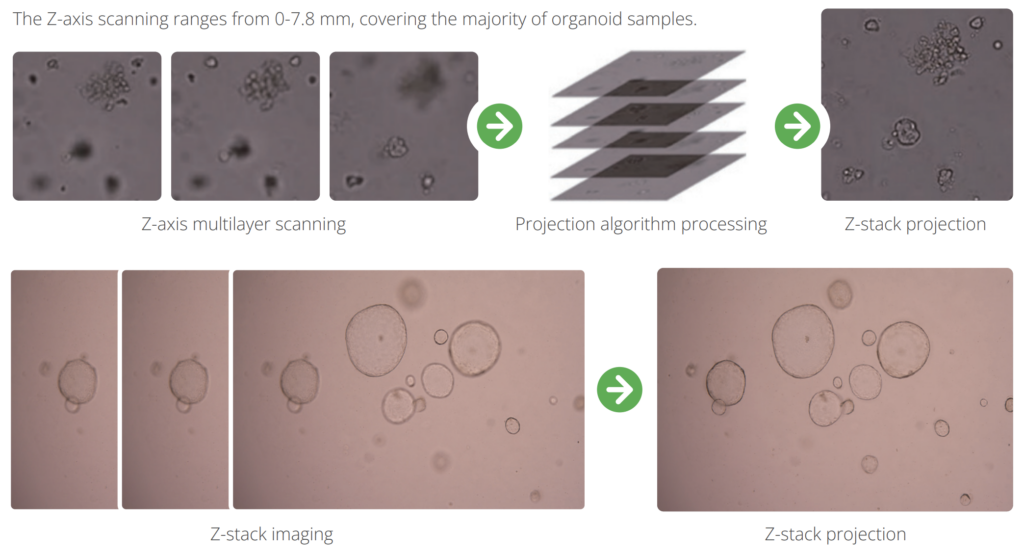
On the Castor S1, the ultrafast Z-axis scanning function addresses a major challenge in high-throughput 3D image acquisition. Clear 2D images are captured along the Z axis at ultra-high speed, dramatically reducing the image acquisition time. It takes only 4 min to complete image acquisition for a 96-well plate when ten 2D images are acquired per well. The linked images of each well are projected to construct a 2D image where cells or organoids at different focal lengths are shown in focus, enabling a comprehensive analysis of each organoid’s phenotype characteristics or confident identification of single cells in monoclonality assay.
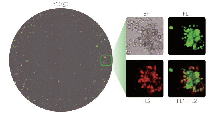
Equipped with red/green dual fluorescence channels, Castor analysers facilitate many kinds of dual fluorescence assays to accomplish your research goals.
The pre-installed consumable library contains the design data of a wide range of consumables from various suppliers. New consumables can be easily added to the library by the user.
Powerful data management and control capabilities make Castor analysers fully compliant with FDA 21 CFR Part 11 regulations.
The development of innovative drugs or cell-based therapies demands significant higher throughput in cell-based experiments. Castor X1 is the ideal platform for many high throughput cell-based assays, including clone identification, confluence, viability and cell transfection analysis.
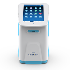
Castor X1
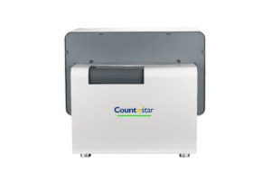
Castor S1
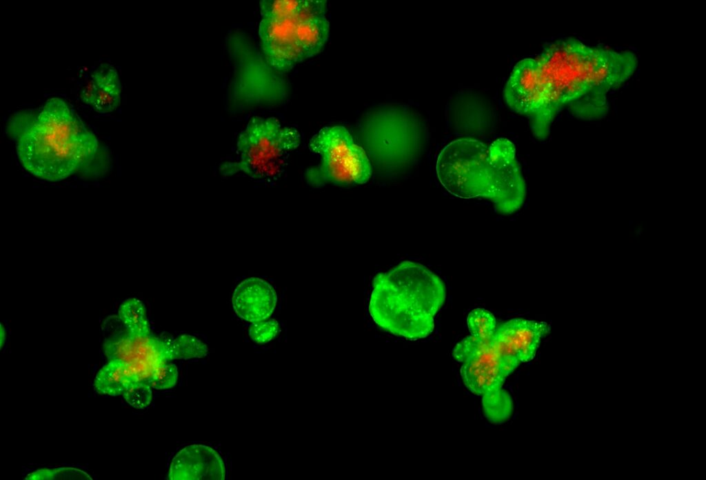
With the emergence of organoids as a potential alternative to model human tissue, interest in how these three-dimensional (3D) structures reacts to its environment is growing among the research and biopharmaceutical community. The development of innovative organoid based research or organoid-based therapies demands accurate and significant higher throughput organoid-based experiments that can accurately capture the physiology of these 3D structures. Castor S1 is the ideal platform for many high throughput organoid-based assays, including features to perform tailored applications such as organoid culture quality, drug-response test, organoid viability, and cytotoxicity analysis for co-culture assay with immune cells.
| Models | Castor X1 | Castor S1 |
|---|---|---|
| Image analysis | 2D | 3D |
| Light source | High performance LEDs | |
| Optical magnifications | 4x objective lens | 4x objective lens (NA=0.2) 10x objective lens (NA=0.3) |
| Imaging elements | 16-bit, 10-megapixel, cooled CMOS colour camera | |
| Focus mode | Laser and image-based autofocus | |
| Compatible consumables | From 6- to 384-well plates Microscope slides 35, 60 and 100 mm petri-dishes T25 and T75 flasks | |
| Fluorescence channels: Excitation wavelengths (nm) Emission filters (nm) | 480/30, 540/25 535/40, 620/60 | |
| Scope of Z-axis scanning | N/A | 0 - 7.8 mm |
The Countstar Castor X1 is the ideal platform for many high throughput assays, including monoclonal identification, cell transfection analysis, virus titre detection and convergence analysis.
The number of cells in each well is counted and tracked automatically throughout the cell culture duration. The AI powered algorithm ensures accurate identification of monoclonality for both adherent and suspended cells, reducing manual verification time by 65%.
With dual fluorescence channels, cells transfected with GFP and/or RFP can be accurately identified and quantified, ensuring confident evaluation of transfection efficiency in cell and gene therapies, cell line development for antibody drugs.
Viral titre is calculated by counting the number of plaques formed on monolayer cells after virus infection.
High-throughput cell convergence analysis can be used in the assessment of cell proliferation, migration, cytotoxicity and cell culture quality control. It is also the foundation of normalising antibody expression level in cell transfection and other downstream analysis.
The Countstar Castor S1 is the ideal platform for many high-throughput organoid-based assays, including features to perform tailored applications, such as organoid culture quality, organoid viability and cytotoxicity analysis for co-culture assay with immune cells.
Castor S1 automatically tracks each individual organoid in the entire culture well plate throughout the organoid culture duration. Its Z-stacking feature and AI powered algorithm enables comprehensive phenotype analysis of each organoid.
Castor S1’s innovative optical design allows capturing bright and clear images of the entire well area in a culture plate. Its AI powered algorithm and automated data analysis ensures accurate and high throughput measurement of organoids morphology during their growth time in each individual cells in the culture plate.
With bright field imaging capability and dual fluorescence channels, cell viability in organoids and tumour spheroids can be measured using viability staining reagents (e.g. Acridine Orange / Propidium Iodide). Fluorescence imaging in a Z-axis scan is carried out to obtain the titre, distribution, and spatial localisation of dead and live cells. This is essential in evaluating the tissue penetration effect of drugs.
Patient-derived organoids are a powerful and effective model for drug screening and patient response prediction to treatment regimens, especially with immune cells co-culture. Castor S1 enables high-throughput imaging and effective data processing, accelerating the identification of an optimal, individualised treatment for the patients.