


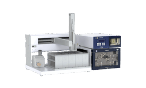


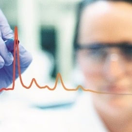


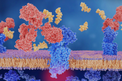


Next-generation label-free analysis
BLI is a powerful technology that is flexible, highly-sensitive and accessible. From experiment to analysis, characterise the widest range of molecules on a single easy-to-use platform ― in a single run.
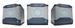
True low volume, high throughput subvisible particle analysis
Aura systems help you develop stable, pure, and efficacious protein, cell, and gene therapies faster. By combining the sensitivity of BMI with fluorescent detection, Aura systems enable high-throughput particle count, size and identification.
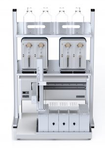
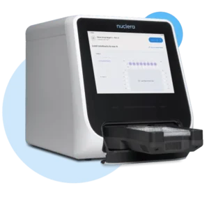
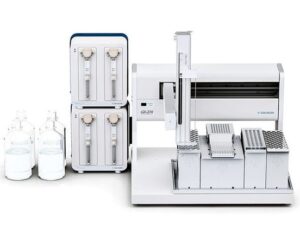
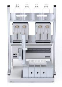
Rapid protein prototyping – Express protein on your own terms
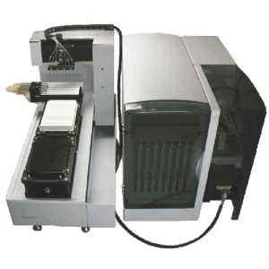
Nuclera’s eProtein Discovery is a benchtop system combining digital microfluidics alongside in situ protein detection assays and cell-free protein synthesis. The eProtein Discovery™ solution, including integrated eProtein Discovery Software, gets protein in your hand quicker than ever before. With our cell-free protein synthesis reagents and microfluidics-based eProtein Discovery™ cartridges, it all happens on your own benchtop, in your way.
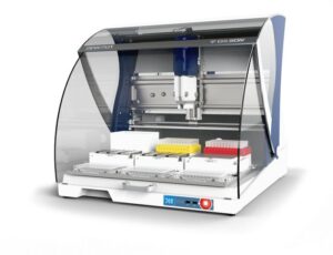
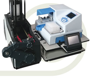
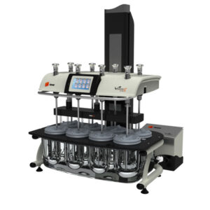
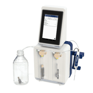
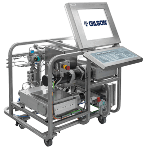
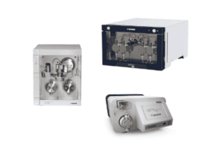
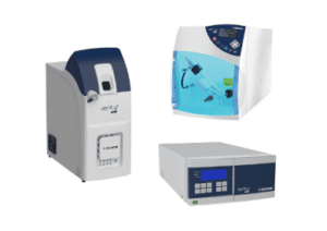
Advancing bioprocessing with precision & performance
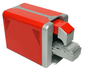
Explore Sepax’s portfolio where innovation meets reliability. Empower your processes with cutting-edge solutions designed for excellence in biomolecule separation and purification.
Next-generation label-free analysis
BLI is a powerful technology that is flexible, highly-sensitive and accessible. From experiment to analysis, characterise the widest range of molecules on a single easy-to-use platform ― in a single run.
True low volume, high throughput subvisible particle analysis
Aura systems help you develop stable, pure, and efficacious protein, cell, and gene therapies faster. By combining the sensitivity of BMI with fluorescent detection, Aura systems enable high-throughput particle count, size and identification.
Rapid protein prototyping – Express protein on your own terms
Nuclera’s eProtein Discovery is a benchtop system combining digital microfluidics alongside in situ protein detection assays and cell-free protein synthesis. The eProtein Discovery™ solution, including integrated eProtein Discovery Software, gets protein in your hand quicker than ever before. With our cell-free protein synthesis reagents and microfluidics-based eProtein Discovery™ cartridges, it all happens on your own benchtop, in your way.
Advancing bioprocessing with precision & performance
Explore Sepax’s portfolio where innovation meets reliability. Empower your processes with cutting-edge solutions designed for excellence in biomolecule separation and purification.
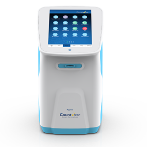
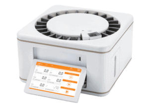
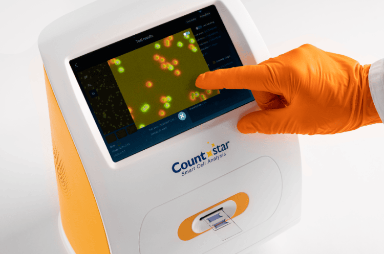
The Countstar Mira systems are automated cell counters that provide exceptional accuracy, sensitivity and reproducibility for a broad range of cell analyses. The Mira counters integrate artificial intelligent (AI) algorithm, patented fixed-focus technology and advanced optical technology to elevate and simplify cell counting and cell analysis for a variety of cell types for users of all levels.
The Countstar Mira cell counters come with fixed focus technology to eliminate errors associated with manual focusing or auto focusing. This dramatically reduces analysis time while ensuring the consistent capture of clear high-resolution images. Using Countstar’s 5-Chamber Slides, the Miras analyse 5 individual samples in a single sequence, assuring highly precise and accurate results while saving time and optimising workflow.
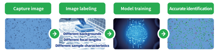
The Countstar Mira software allows user friendly operation while also providing the technical adeptness of distinguishing various cell types with irregular morphology, clustering, while excluding non-desirable particles and debris. The Artificial Intelligence (AI) based, self-learning image analysis algorithms are capable of accurately identifying and quantifying small aggregated suspension cells, organoids, and a wide variety of adherent cells.
The advanced AI based image analysis algorithms of the Mira deliver all essential cell viability parameters to evaluate the actual status of a cell culture. Along with cell density and viability, the cell size distribution, the cell shape factor, and any formation of cell clusters (aggregates) are analysed as well. The relative fluorescence intensity delivers you data about the content of markers or fluorescently tagged cells. The growth curve feature allows an immediate feedback about the kinetics of a running bio-process. Single cells can be classified even inside aggregates. Changes of the cell shape factor can be an important, early-stage indicator for changes in your cell culture. The data provided from the Mira enables better interpretation for users to optimise the dynamics of cell culture processes with mammalian, algae, and yeast cells.
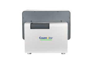
The Mira BF automated cell analyser integrates advanced optical imaging technology, intelligent image recognition and user-friendly consumable design to achieve the detection and analysis of multiple samples. Results are generated in seconds and provide you with a full range of cell culture information that may assist in determining ideal cell culture conditions.
Additionally, Mira BF can function as a digital microscope to image culture slides, flasks and petri dishes, expanding it’s functionality to provide confluence and organoid analysis.
Mira BF
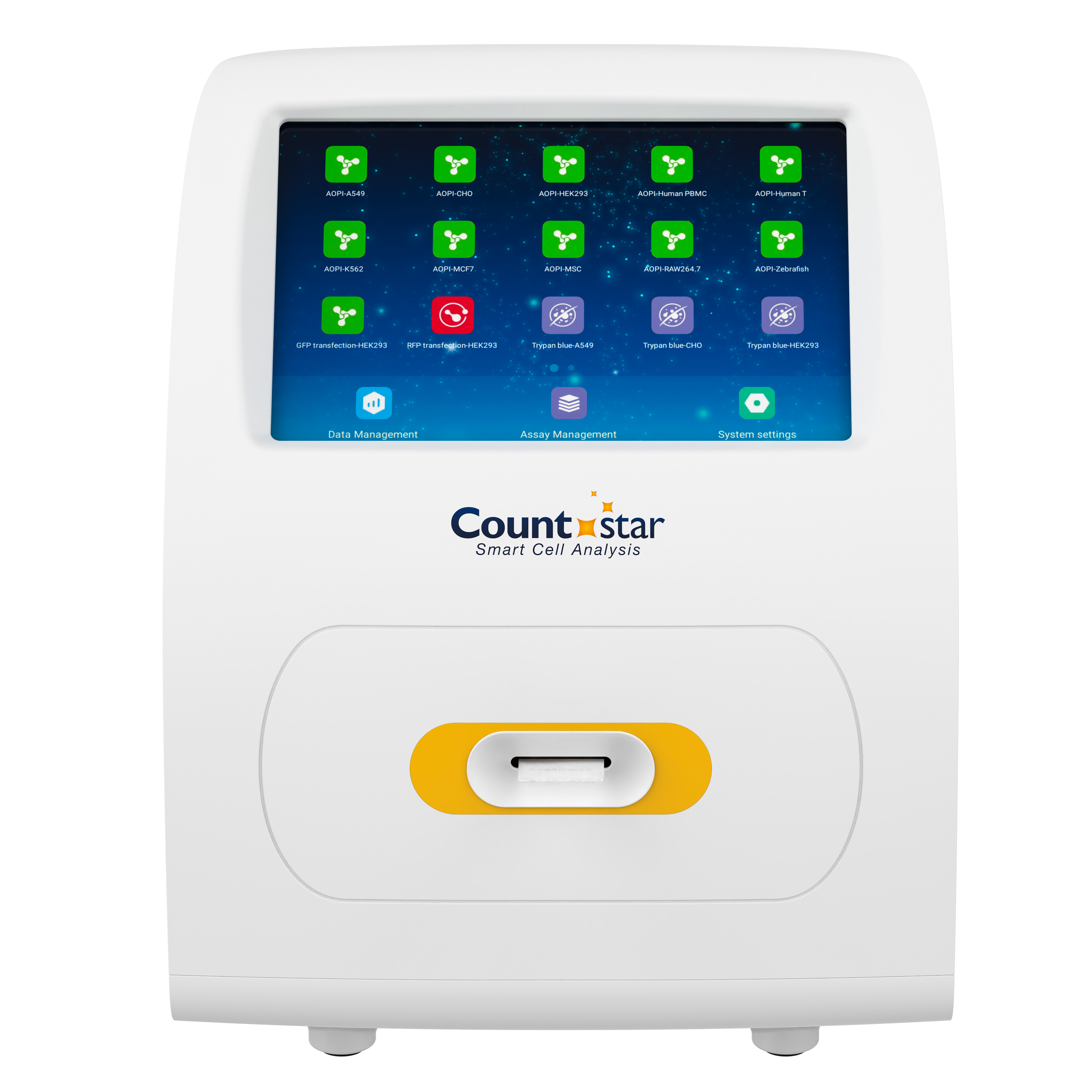
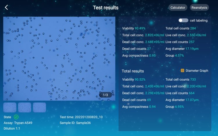
The Mira FL cell analyser integrates AI intelligent algorithm and adopts patented fixed focus and optical zoom technology to realise the identification of cell characteristics. With trypan blue and AO/PI staining methods, it helps to achieve accurate counting of all types of cells and supports GFP/RFP transfection experiments. The instrument is easy to operate, efficient in analysis and testing, saves valuable scientific research time, and helps scientific research laboratory personnel to achieve fast and efficient cell analysis result.
Mira FL
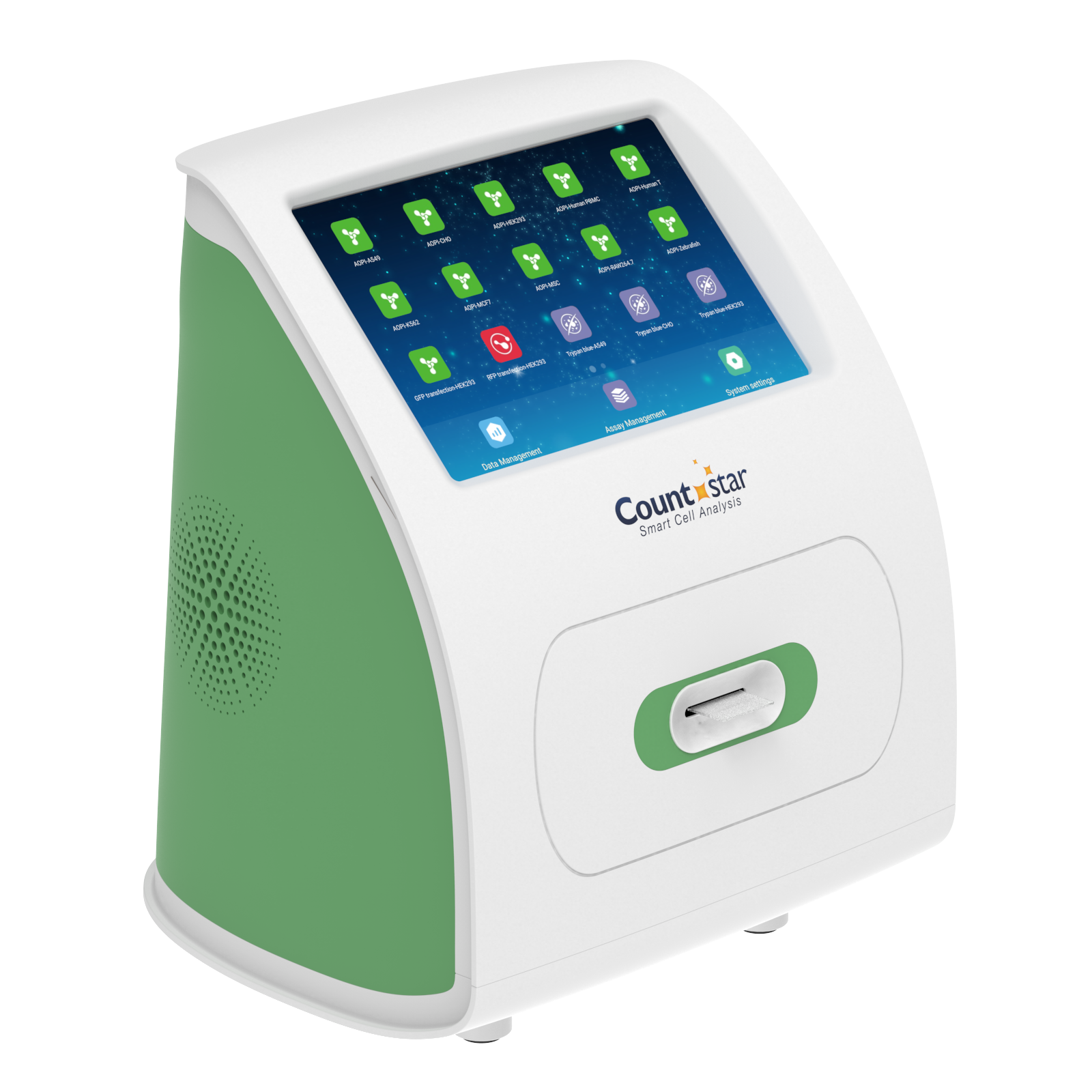
The Mira FL Plus is an automated fluorescence cell analyser that provides exceptional accuracy, sensitivity and reproducibility for a broad range of cell analyses. Mira FL Plus has advanced high-definition optics to capture high-resolution images, and a larger field of view for higher precision cell counting.
Mira FL Plus is fully compliant, meeting the regulatory requirements of FDA 21 CFR Part11 and cGMP.
Mira FL Plus
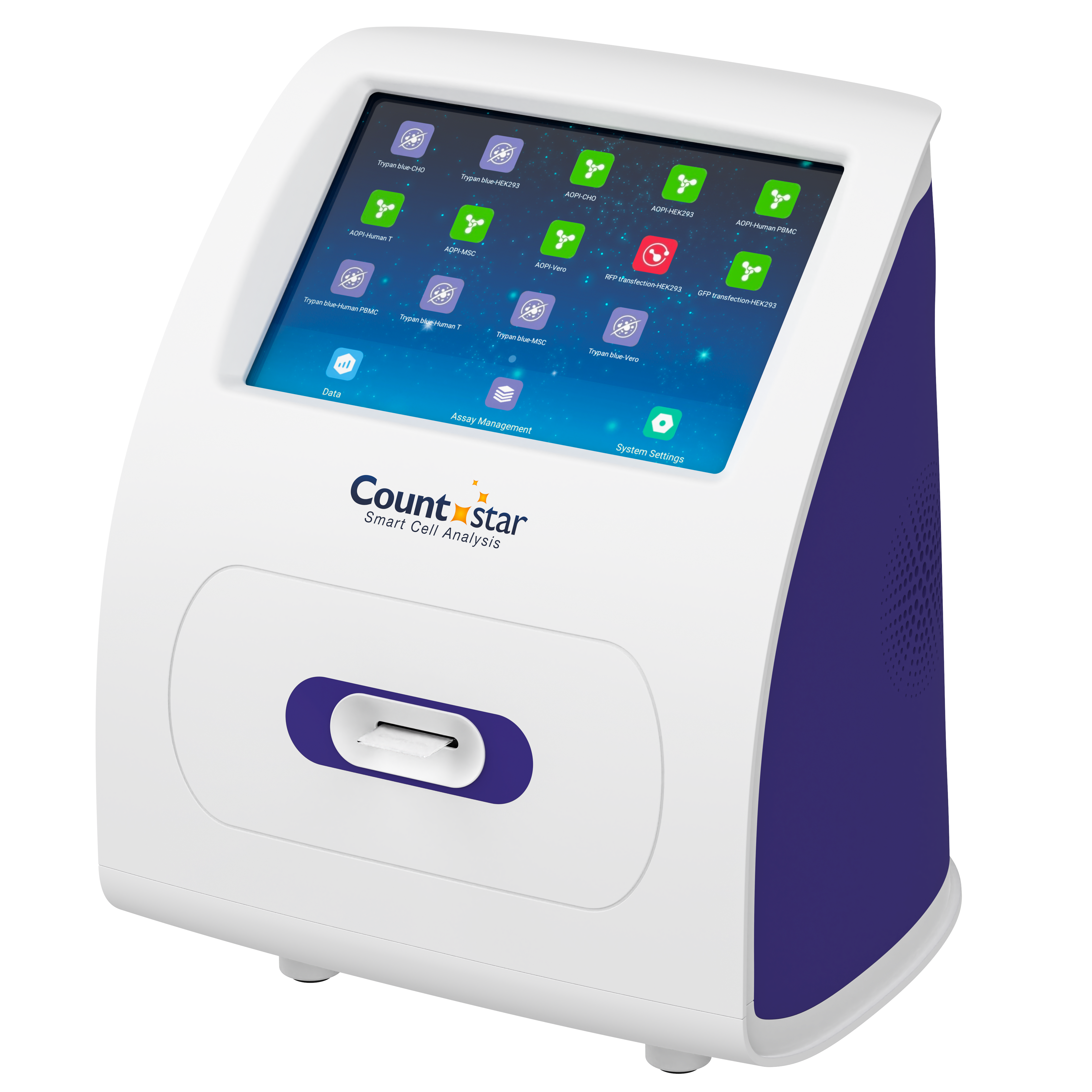
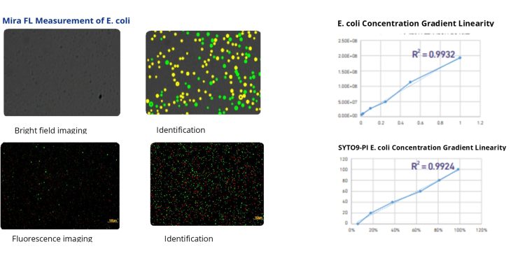
The Mira FL Pro microbial cell analyser uses 10x optical magnification, 8.3 million CMOS camera and innovative “fixed focus” technology to achieve high quality images of small cells such as microorganisms. Mira FL Pro can detect objects in a size range from 1 – 10 μm. With AI intelligent learning algorithms, based on rich sample database model training, Mira FL Pro ensures the accuracy and stability of the data.
Mira FL Pro is fully compliant, meeting the regulatory requirements of FDA 21 CFR Part11 and cGMP. It can meet different experimental requirements such as precise cell counting, viability detection and fluorescence analysis.
Mira FL Pro
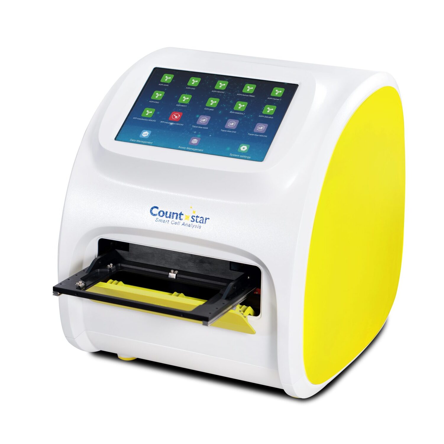
Building off the previous Mira platforms, the Countstar Mira HT performs both brightfield and fluorescence analysis but with the capabilities of high-throughput sampling in a compact package. Also unique is the versatility and range of assays the Countstar Mira HT is capable of performing, such as AO/PI viability and GFP/RFP transfection detection.
Mira HT is fully compliant, meeting the regulatory requirements of FDA 21 CFR Part11 and cGMP.
Mira HT
| Mira models | BF | FL | FL Plus | FL Pro | HT |
|---|---|---|---|---|---|
| Cell counting throughput | 5 | 5 | 5 | 5 | 15* / 24~ |
| Consumable compatibility | 5-Chamber slide Petri dish T25 culture flask Haemocytometer Chamber culture slide | 5-Chamber slide | 5-Chamber slide 24-Chamber plate | ||
| Optical magnification: Brightfield Fluorescence | 5.0x, 6.6x, 8.0x N/A | 5.0x, 6.6x, 8.0x 5.0x | 5.0x 5.0x | 10.0x 10.0x | 5.0x 5.0x |
| Cell types | Mammalian cells Yeasts and algae Organoids / spheroids | Mammalian cells Yeasts and algae | Mammalian cells | Bacterial cells Yeasts and algae Platelets | Mammalian cells |
| Cell detection size range (μm) | 2 – 180 | 1 - 180 | 1 - 10 | 2 - 180 | |
| Concentration range | 1 × 10⁴ - 3 × 10⁷ cells / mL | 5 × 10⁵ - 2 × 10⁸ cells / mL | 1 × 10⁴ - 3 × 10⁷ cells / mL | ||
| Detection modes | Brightfield | Brightfield and fluorescence | |||
| Fluorescence channels | N/A | Ex.: 465 - 485 nm Em.: 535 / 40 nm; 600 nm LP | |||
| Applications | Trypan Blue Viability Confluence Analysis Object Counting Organoid Analysis | Trypan Blue Viability AO/PI Viability GFP / RFP Transfection Efficiency | Yeast Viability Platelet Viability Bacteria Viability | Trypan Blue Viability AO/PI Viability GFP / RFP Transfection Efficiency | |
| cGMP Compliance | No | Yes | |||
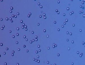
The Trypan Blue based exclusion method is commonly used in many labs to monitor cell culture status and is the gold standard in determination of concentration and viability. Due to the intact membrane structure in living cells, the Trypan Blue dye will be prevented from entering a cells cytoplasm. Dying cells that lose activity and whose membranes get disintegrated cause a permeability for the stain Trypan Blue.
This optical appearance of living and dead cells can be precisely discriminated using the AI based image analysis algorithms of the Mira cell counters. Beyond cell number of dead and viable cells as results, Mira’s image analysis delivers additional data about the cells in focus: Cell diameters, aggregate formation, and cell morphology.
The Countstar Mira BF automated cell analyser features the ability to analyse the confluency of adherent cells grown in petri dishes and T25 flasks. Using innovative AI-based image recognition algorithms, the adherent cell area and the non-adherent single cells are marked to obtain the confluency and adherence rate simultaneously. The evaluation of the cell proliferation by generating a growth curve in the software provides a new method for precise and accurate assessment of different cell lines.
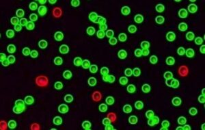
The AO/PI dual dye fluorescence BioApp for determination of cell density and viability of eukaryotic cell cultures is based on the principle that both dyes, Acridine Orange (AO) and Propidium Iodide (PI), will intercalate between the nucleic acids of the chromosomes inside the nucleus. AO will permeate an intact nucleus membrane at any time to stain the DNA, emitting a green light at 525nm max., if excited at 480nm. PI is only capable to pass a disintegrating nucleus membrane of a dying (dead) cell. This dye will emit a red light with its amplitude at 615nm, if PI is excited at 525nm.
The AO/PI staining kit utilises the physicochemical phenomenon of the FRET-effect (Foerster Resonance Energy Transfer). If samples of cells are excited at 480nm, the emitting light of the present AO dye in the nucleus at 525nm will be totally absorbed by the accumulated PI dye in the nuclei of dead (dying) cells, which then emit due to their excitation the red light at 615nm. The energy transfer ensures that no double light emission or overexposure of light will occur. Additionally, it also ensures that only nuclei containing cells will get stained and no akaryotes, like erythrocytes, will be included.
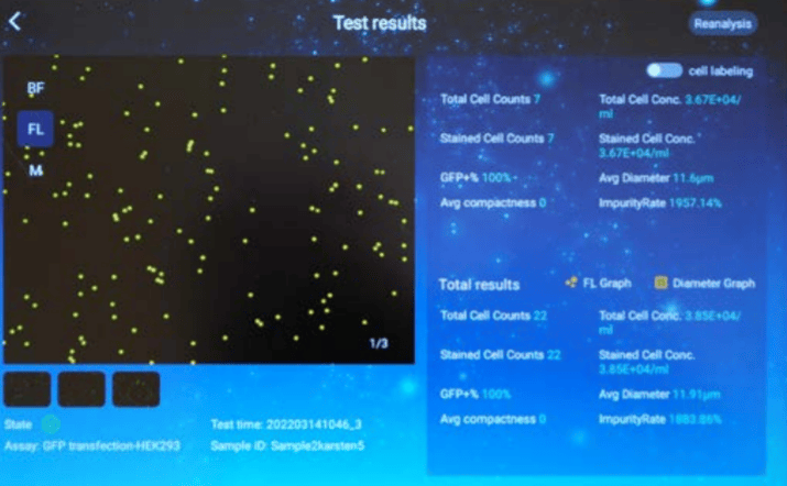
Using green or red fluorescence proteins (GFP / RFP) to bind them covalently as marker tags to the protein of interest, is a widely established technique to check the transfection efficiency of viral or plasmid vector-based genes. GFP/RFP are reliable tools to quantify the optimisation of a protein manufacturing process, and is a fast, and highly precise method to determine the protein yield in many cGMP regulated manufacturing processes of APIs. This labelling technology has also become an important tool in various gene therapeutic approaches to control the transfection efficiency of genetic modifications.
Compared to flow cytometry, Mira does not only deliver comparable results, but provides additional images to validate the results of questionable cells. All images and data are permanently stored, offering a high level of traceability. Additionally, imaged based analysis is significantly faster, and needs only 20 µL of sample liquid. The Mira simplifies this analysis workflow, helping to streamline each transfection optimisation process.
The Mira FL Pro provides high-definition imaging and accurate algorithmic identification to help you achieve accurate counts of small cells such as platelets, yeasts and bacteria. Mira FL Pro is equipped with both brightfield and red & green fluorescence channels, expanding it’s applications to assess viability via fluorescence staining of yeast (AO/PI), platelets (Calcein-AM) and bacteria (Styo-9/PI).