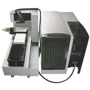


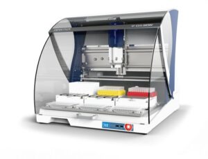


Next-generation label-free analysis
BLI is a powerful technology that is flexible, highly-sensitive and accessible. From experiment to analysis, characterise the widest range of molecules on a single easy-to-use platform ― in a single run.
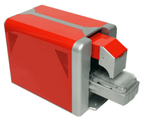
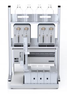
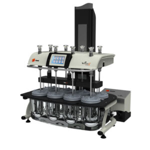
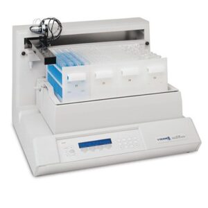
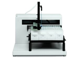
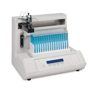
True low volume, high throughput subvisible particle analysis
Aura systems help you develop stable, pure, and efficacious protein, cell, and gene therapies faster. By combining the sensitivity of BMI with fluorescent detection, Aura systems enable high-throughput particle count, size and identification.
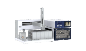
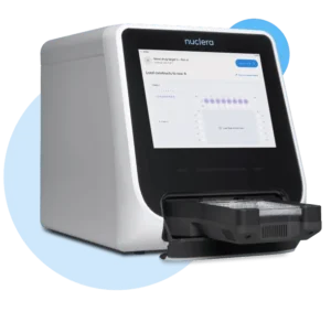
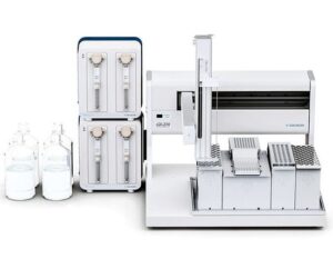
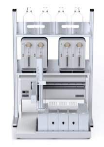
Rapid protein prototyping – Express protein on your own terms
Nuclera’s eProtein Discovery is a benchtop system combining digital microfluidics alongside in situ protein detection assays and cell-free protein synthesis. The eProtein Discovery™ solution, including integrated eProtein Discovery Software, gets protein in your hand quicker than ever before. With our cell-free protein synthesis reagents and microfluidics-based eProtein Discovery™ cartridges, it all happens on your own benchtop, in your way.
Next-generation label-free analysis
BLI is a powerful technology that is flexible, highly-sensitive and accessible. From experiment to analysis, characterise the widest range of molecules on a single easy-to-use platform ― in a single run.
True low volume, high throughput subvisible particle analysis
Aura systems help you develop stable, pure, and efficacious protein, cell, and gene therapies faster. By combining the sensitivity of BMI with fluorescent detection, Aura systems enable high-throughput particle count, size and identification.
Rapid protein prototyping – Express protein on your own terms
Nuclera’s eProtein Discovery is a benchtop system combining digital microfluidics alongside in situ protein detection assays and cell-free protein synthesis. The eProtein Discovery™ solution, including integrated eProtein Discovery Software, gets protein in your hand quicker than ever before. With our cell-free protein synthesis reagents and microfluidics-based eProtein Discovery™ cartridges, it all happens on your own benchtop, in your way.
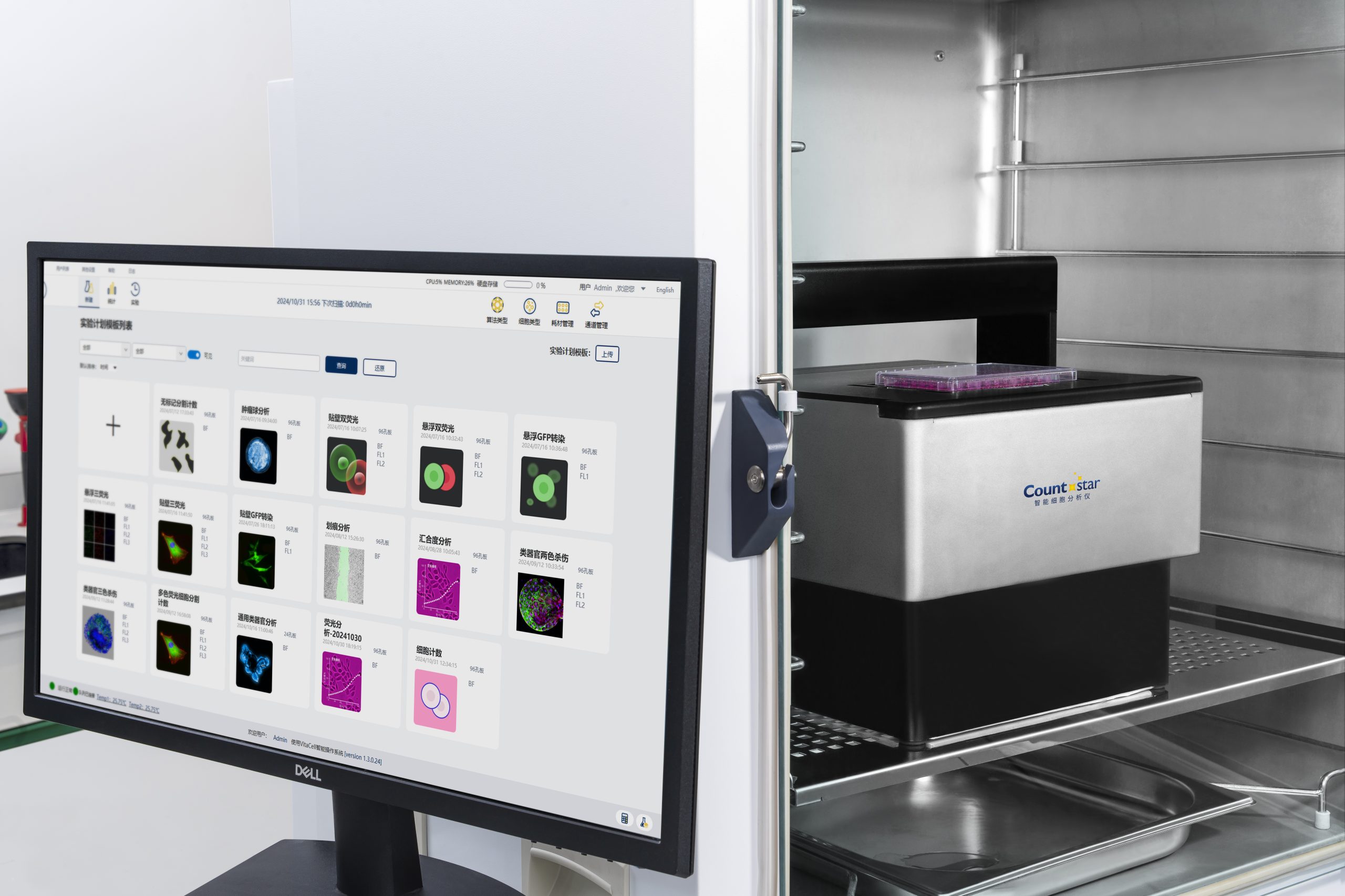
In the dynamic world of cell biology research, the ability to adapt and explore diverse scientific questions is paramount. Countstar’s goal is to empower biological researchers worldwide to explore the dynamic world of living cells with advanced live cell imaging and analysis tools. Countstar’s Spica systems have built-in flexibility to enable seamless transitions between different assays, allowing investigation of multiple cellular processes with existing tools in your own laboratory.
Incubator-friendly for optimal cell health
Countstar’s Spica systems fit directly inside any standard incubator, ensuring physiological conditions for reliable long-term imaging.
Ready-to-go assays
Start experiments faster with preset parameters for common applications and ensure consistent, reliable data for all users.
Seamless imaging to analysis, all in one
Integrated software streamlines your entire workflow, from acquisition to visualisation. No need for multiple programs or extra costs.
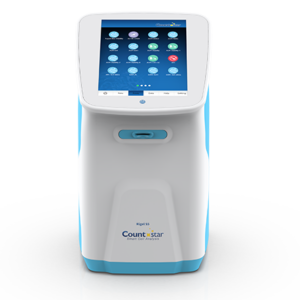
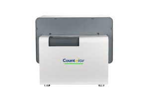
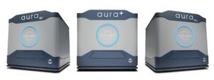
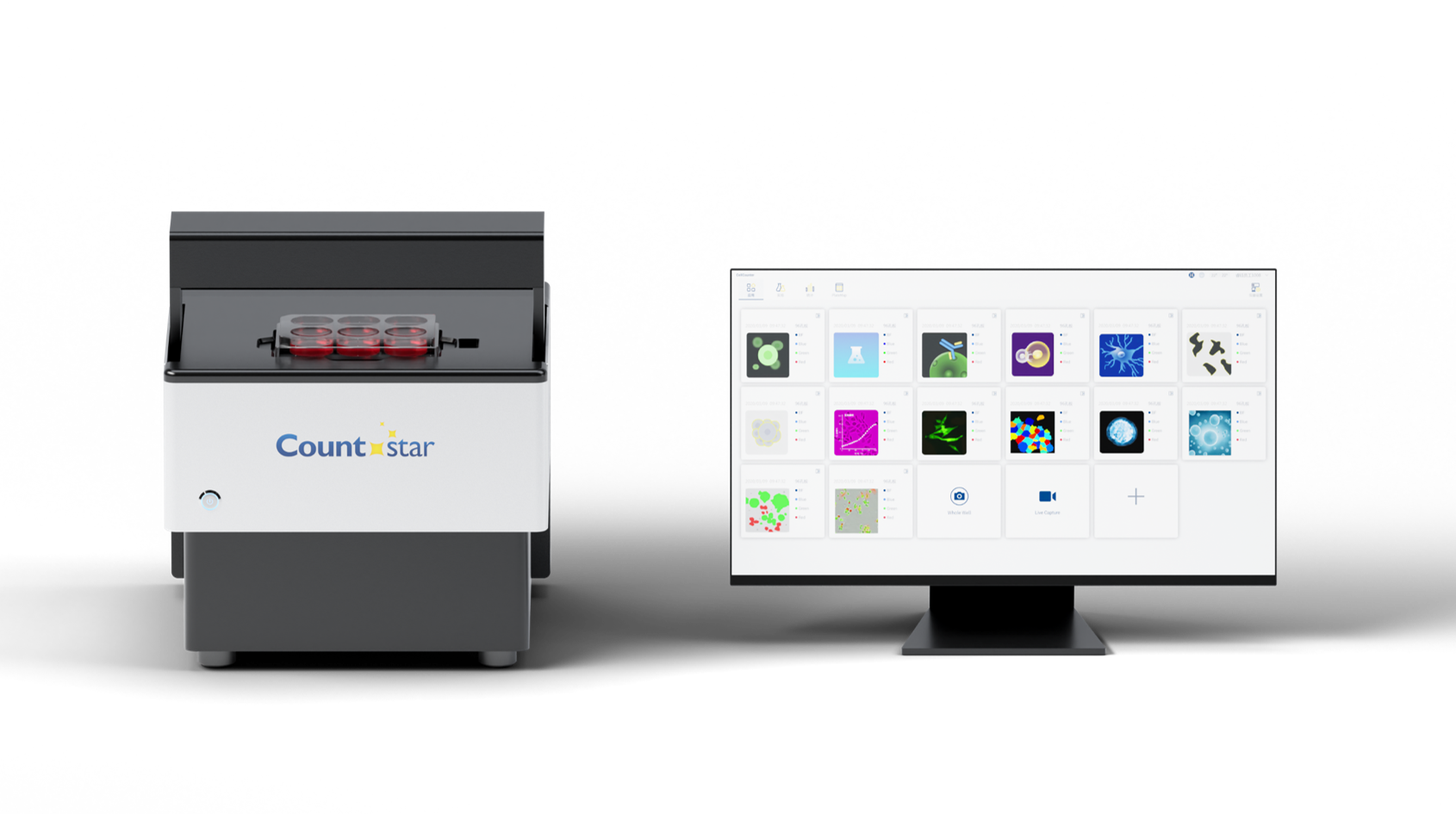
Spica M1 live cell analysers
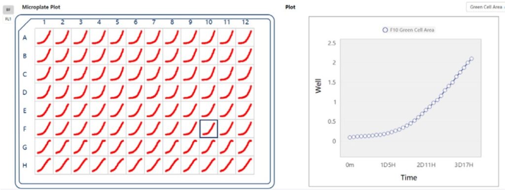
Spica allows you to stay connected to your ongoing experiments and track critical cellular events remotely, increasing flexibility and efficiency.
Spica’s integrated AI analysis tools efficiently process various datasets to uncover insights. They enable quick identification of key features and automated quantification, even in complex experiments that traditional methods struggle to analyse.
Spica allows you to customise imaging parameters and analysis metrics to meet the unique demands of your diverse assays, providing the exact data you need.
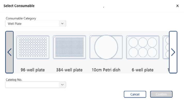
Image various cell types and markers simultaneously with multiple channels, observe at different scales with flexible magnification and utilise your preferred lab consumables.
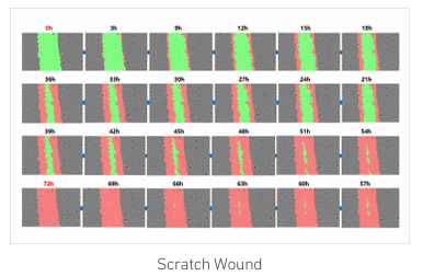
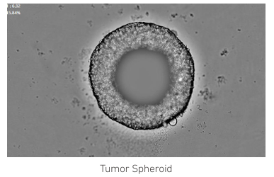
Live Capture Mode allows easy observation and recording of dynamic, short-lived cellular processes in real time, providing immediate insights into critical biological events.
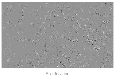
High resolution brightfield and fluorescence live cell imaging, with intelligent and flexible analysis for research
2 fluorescent channels (red & green)
3 fluorescent channels (red, green & blue)
Spica M1 live cell analysers
Real time live cell analysis
| Spica Models | M1-1 | M1-2 |
|---|---|---|
| Plate capacity | 1 | 1 |
| Microscope objectives | 7.5 x, 10 x and 15 x | |
| Fluorescence channels: | 2 channels: Green Ex: 470/20 Em: 515/40nm Red Ex: 560/25 Em: 630/70nm | 3 channels: Blue Ex: 375/28 Em: 430/30nm Green Ex: 470/20 Em: 515/40nm Red Ex: 560/25 Em: 630/70nm |
| Camera | High-sensitivity CMOS camera, 3200 × 2200, 7.1MP | |
| Focus mode | Laser autofocus and image-based autofocus | |
| Imaging modes | Bright-field and fluorescence microscopy Whole well scanning, fixed fields and random fields of view 2D and 3D Z-axis layer scanning | |
| Compatible consumables | 6 - 384 well plates 35 mm, 60 mm and 100 mm dishes T 25 and T 75 flasks Organ-on-a-chip microfluidic chips | |